Showing posts with label PATHOLOGY. Show all posts

Friday, 20 May 2011

Atlas of Pathology images of Fibrocystic disease changes

A mass characterized by fibrocystic change is a pathological rather than a clinical diagnosis. A fibrocystic mass is diagnosed based on the presence of 2 important features: fibrous connective tissue and cysts

Fibrocystic changes are defined as Cystic changes and apocrine metaplasia involving terminal duct lobular units (TDLU) of breast tissue.

This image shows Extensive fibrocystic changes in serially sectioned formalin fixed breast tissue :
Note that the cysts are of various size are interspersed by dense fibrous tissue. This patient, who was in a high risk category for breast carcinoma, elected to have bilateral mastectomies with prosthetic implants. This degree of "fibrocystic change" becomes impossible to follow on mammograms due to complex shadows from the dense fibrous tissue.

Another gross picture for breast tissue with fibrocystic changes :


Note presence of "Blue dome" wich is a benign cysts filled by serous fluid often have this blue color when viewed from the outside


The next image is for Microscopic picture of Fibrocystic changes, including cystic dilatation, apocrine metaplasia, florid ductal hyperplasia, and fibrosis.


Monday, 4 April 2011

Gross pathology of Atherosclerosis in Aorta

Atherosclerosis is the process in which deposits of fatty substances, cholesterol, cellular waste products, calcium and other substances build up in the inner lining of an artery "Plaque".

Distribution of atheromatous plaques:
  •  Lower descending aorta {ostia of the major branches}
  •  First 3 inches of coronary arteries
  •  Descending aorta
  •  Internal carotids
  •  Circle of Willis
  •  Leg arteries
This image shows Atheroscleosis of Aorta; the aorta was opned along the posterior wall . Numerous calcified and ulcerated, yellow atherosclerotic plaques (arrows) dot the inner surface. Note the narrowed aorta above the iliac bifurcation. It is calcified, stenotic and lined by clotted blood.

Clinical Consequences of Atheromatous plaques include:
* Occlusion
* Vessel rupture
* Aneurysmal dissection

Saturday, 2 April 2011

Monday, 21 March 2011

Gross and Microscopic picture of Renal Infarction

1- Gross specimen of Renal infarction:

The kidney is cut in half along its longitudinal axis, exposing :
(A.) the cortex
(B.) the medulla
(C.) a minor calyx .
The pyramidal shaped infarct is pale as compared to the adjacent normal cortex. Why? as the wedge shape of this zone of coagulative necrosis resulting from loss of blood supply with resultant tissue ischemia that produces the pale infarct.
The arrow points to a line of hyperemia that represents the interface between normal and necrotic tissue.

2- Microscopic section of Renal infarction:

* The thick arrow points to glomerulus in an area of coagulation (ischemic) necrosis.
* The thin arrow points to a glomerulus which is in the interface between necrotic and normal kidney.

Wednesday, 29 December 2010

Benign Prostatic Hyperplasia

Many men with benign prostatic hyperplasia experience urinary problems related to the condition. As the prostate enlarges, the gland places increasing pressure on the urethra, often resulting in difficulty beginning or ending urination, an inability to completely empty the bladder, decreased urine flow, and frequent urination. In the most severe cases, complete blockage of the urethra occurs, which may lead to kidney damage.
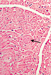
From microscopyu.com

Benign Prostatic Hyperplasia at 20x Magnification :
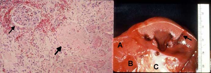
Part of the male reproductive system, the prostate gland produces and stores seminal fluids, releasing them into the urethra when semen emission occurs. The gland is located directly below the bladder and surrounds the upper part of the urethra. During adolescence the gland usually matures and reaches a size comparable to that of a walnut. The dimensions of the gland generally remain unchanged for several decades, but in most older men, the prostate begins to enlarge as the size of its cells increases, a process commonly referred to as benign prostatic hyperplasia (BPH) or hypertrophy. According to recent estimates, more than 50 percent of men between the ages of 50 and 60 experience benign prostatic hyperplasia, and over 90 percent of those 70 to 90 years old have developed the condition. Researchers do not yet completely understand the cause of this physiological change, but it is widely thought that elevated levels of the female sex hormone estradiol and increased manufacture of dihydrotestosterone, a derivative of the male sex hormone testosterone, contribute to the condition.

Benign Prostatic Hyperplasia at 4x Magnification :
Men with only mild symptoms of benign prostatic hyperplasia may elect not to undergo any treatment or to simply take a wait-and-see attitude, visiting the doctor regularly for monitoring until signs suggest a more active approach is needed. For those who seek treatment, a number of options are available. For example, drugs such as alpha blockers and finasteride may be used alone or in conjunction with one another to relax prostatic smooth muscle and decrease the size of the prostate gland. Individuals that are not responsive to the typical medications, however, may require a more invasive form of treatment, such as balloon dilation of the urethra or any of several different surgical techniques, including transurethral incision of the prostate (TUIP), transurethral resection of the prostate (TURP), or open prostatectomy. The various treatments for benign prostatic hyperplasia are associated with a number of risks and side effects, which can include serious conditions like incontinence and impotence.

Thursday, 23 December 2010

Toxoplasmosis in Immune-Suppressed Patients

Toxoplasma encephalitis (inflammation of the brain) and Toxoplasma myocarditis (inflammation of the heart) are well recognised opportunistic infections in patients who are immune suppressed, particularly in relation to AIDS and chemotherapy for cancer. The Toxoplasma encephalitis has the usual appearance of an encephalitis from any cause, that is, focal areas of death of cerebral tissue associated with a mononuclear inflammatory cell infiltrate. But in addition, Toxoplasma cysts are found in the affected brain tissue.

This figures are from the heart of a middle aged male who died from AIDS. The Toxoplasma cyst is expanding the myocardial muscle fibre. In this case there is no inflammatory reaction associated with the cyst. The presence of an inflammatory reaction is variable.

Wednesday, 22 December 2010

Fibroadenoma

Fibroadenomas, which are generally firm, smooth, and round, can be readily moved under the skin and are often described as feeling similar to marbles. Typically the masses measure from 1 to 3 centimeters in size, but occasionally they may grow much larger, in which case they are termed giant fibroadenomas. Fibroadenomas, which arise from the intralobular stroma, are solid and consist of a combination of glandular and fibrous tissues. The tumors are usually painless and present no symptoms, typically being discovered by young women only due to self-examination. In older women, fibroadenomas are often less palpable and may first be discovered during a routine mammogram.

Fibroadenomas showing circumscribed margins, even distribution of epithelial and stromal components and low stromal cellularity.
-Low power scanning of Fibroadenoma :
# Use low power scanning to determine:
  •  the basic pattern - pericanalicular or intracanalicular
  •  edge of lesion (pushing or infiltrative) - should be pushing
  •  balance between stroma and epithelium - should be even
  •  to pick out areas of stromal hypercellularity


# The stroma can be cellular particularly in younger patient's lesions - but it's usually uniform

# The occasional stromal mitosis is acceptable in a younger patient's lesion but take advice

# Uneven stromal cellularity in a core biopsy may be a pointer to a Phyllodes Tumour

You can suspect Phyllodes Tumour if:
* Patient older than 40 years
* Lesion larger than 4 cm
* History of recent growth