Wednesday, 25 May 2011

Causes, Symptoms and sings of aortic valvular insufficiency

A 74-year-old man presents with exertional dyspnea and generalized weakness. On examination, you discover a high-pitched, blowing diastolic murmur and a wide pulse pressure with bounding pulses. The most likely diagnosis is?
  • A) aortic stenosis
  • B) aortic insufficiency
  • C) mitral stenosis
  • D) mitral insufficiency
  • E) coarctation of the aorta

Answer and Discussion
The answer is B.
Causes of aortic regurgitation :
The prevalence of aortic regurgitation increases with age. Unlike aortic valve stenosis, aortic valvular insufficiency is rarely caused by degenerative aortic valve disease. Acute aortic valvular insufficiency may be due to infective endocarditis, aortic dissection, trauma, or rupture of the sinus of Valsalva. Chronic aortic insufficiency can be caused by valve leaflet disease, including rheumatic heart disease, congenital heart disease, rheumatoid arthritis, ankylosing spondylitis, or myxomatous degeneration. Chronic aortic insufficiency may also be caused by aortic root disease secondary to systemic hypertension, syphilitic aortitis, cystic medial necrosis, ankylosing spondylitis, rheumatoid arthritis, Reiter's disease, systemic lupus erythematosus, Ehlers–Danlos syndrome, and pseudoxanthoma elasticum.

Symptoms and sings of aortic valvular insufficiency :
The symptoms of aortic valvular insufficiency are the same in older persons as they are in younger ones. Usually, the main symptoms are related to heart failure, with exertional dyspnea and weakness being common symptoms. In some elderly patients, symptoms of dyspnea and palpitations may be more common at rest than with exertion. Nocturnal angina pectoris, often accompanied by flushing, diaphoresis, and palpitations, may occur; this is thought to be related to the slowing of the heart rate and the drop of arterial diastolic pressure. The classic findings of a high-pitched, blowing diastolic murmur and a wide pulse pressure with an abruptly rising and collapsing pulse should make the diagnosis of aortic valvular insufficiency easily recognized in elderly patients.

Photo description of Pterygium of the eye

Pterygium is a raised, wedge-shaped, non-cancerous growth of the conjunctiva onto the cornea, characterized by elastotic degeneration of and fibrovascular proliferation
Pterygium has a characteristic shape as it is triangular, with the base of the triangle located in the conjunctiva and the apex of the triangle encroaching onto the cornea.
With corneal involvement, even if arrested surgically, a pterygium can affect vision by warping the surface of the cornea and inducing astigmatism, and/or by actually growing over in front of the pupil and obstructing the entering light.
 

Triangular, thin, transparent conjunctival fold in the palpebral fissure. Its head is yellow, avascular and pints toward the corneal center. The body of the pterygium extends to the semilunar fold(this image for a 32-year-old man).

Treatment :
Pterygia seem to occur more frequently in people who spent much time outside, and is especially common in the southern lattitudes "Some consider pterygium as an occupational disease" So protecting the eyes from sun, dust and wind and use of lubricating drops is the standard treatment.
If the pterygium is still troublesome it can be removed surgically under local anaesthetic ,with high rate of recurrence. Once the pterygium is removed, the bare sclera is covered with conjunctiva

Saturday, 21 May 2011

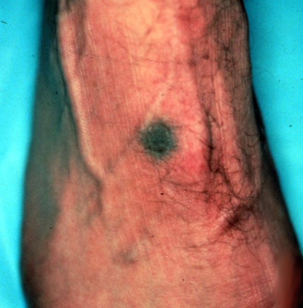
Photos and information about Blue Nevus

- Blue Nevus is a group of melanocytic lesions which all appear blue in colour due to the optical effects of light reflecting off melanin deep in the dermis.

- The blue nevus is a benign, usually solitary lesion, represents a localized proliferation of dermal melanocytes. It presents as a dark blue to black, moderately firm, rounded, sharply defined nodular tumour composed of spindle-shaped melanocytes with slender cytoplasmic processes, occurring often in association with melanin-laden macrophages in a sclerotic dermis.
- A blue naevus (nevus) is a rather unusual but non-cancerous mole.

- The blue nevus is also called the blue skin mole, or Jadassohn-Tièche nevus with 2 clinically recognized variants : the common blue nevus and the cellular blue nevus.
- Cellular blue nevus is larger, especially on buttocks and can degenerate into malignant melanoma.

- Clinically The blue nevus appears as a dark-blue or blue-black smooth nevus formed by melanin-heavily pigmented spindle cells in the middle and lower two-thirds of the dermis. Also, the blue nevus appears as a slate blue or bluish black, sharply circumscribed, flat or slightly elevated nodule, occurring on any area of the body.
- A biopsy should be performed on any changing pigmented lesion. For a solitary lesion, simple excision is usually curative. Rare cases of persistent blue nevi, manifesting as satellite lesions around the original excision site, have been reported. These must be distinguished from malignant blue nevus, and reexcision is recommended.

Common blue nevus

Friday, 20 May 2011

Atlas of Pathology images of Fibrocystic disease changes

A mass characterized by fibrocystic change is a pathological rather than a clinical diagnosis. A fibrocystic mass is diagnosed based on the presence of 2 important features: fibrous connective tissue and cysts

Fibrocystic changes are defined as Cystic changes and apocrine metaplasia involving terminal duct lobular units (TDLU) of breast tissue.

This image shows Extensive fibrocystic changes in serially sectioned formalin fixed breast tissue :
Note that the cysts are of various size are interspersed by dense fibrous tissue. This patient, who was in a high risk category for breast carcinoma, elected to have bilateral mastectomies with prosthetic implants. This degree of "fibrocystic change" becomes impossible to follow on mammograms due to complex shadows from the dense fibrous tissue.

Another gross picture for breast tissue with fibrocystic changes :


Note presence of "Blue dome" wich is a benign cysts filled by serous fluid often have this blue color when viewed from the outside


The next image is for Microscopic picture of Fibrocystic changes, including cystic dilatation, apocrine metaplasia, florid ductal hyperplasia, and fibrosis.