Showing posts with label CHEST. Show all posts

Wednesday, 15 June 2011

Types of respiratory pattern of breathing

A 55-year-old man with emphysema will have which kind of respiratory pattern of breathing?
  • a.Biot respiration
  • b.Apneustic breathing
  • c.Cheyne-Stokes respiration
  • d.Rapid and shallow breathing
  • e.Kussmaul breathing

The answer is d.
In emphysema, there is destruction of alveolar septa and reduced elastic recoil. This causes collapse of the small airways and prolongs the expiratory phase of respiration.
During the prolonged expiration, patients will “purse” their lips to avoid collapse of the small airways. The respiratory rate is increased by having a markedly shortened inspiratory interval.

Kussmaul respirations are slow and deep respirations to increase the tidal volume in patients with diabetic ketoacidosis. Biot respirations are seen in patients with increased intracranial pressure. These are irregular, unpredictable periods of apnea alternating with periods of noisy hyperventilation. Cheynes-Stokes respirationis a rhythmic, gradually changing pattern of apnea and hyperpnea that is cardiac or neurologic in origin. Apneustic breathing is characterized by a long period of inspiration or gasping with almost no expiratory phase.

Saturday, 14 May 2011

Triad of Kartagener syndrome

 A 14-year-old boy presents with a history of chronic sinusitis and frequent pneumonias. On physical examination, the patient has normal vital signs and is afebrile. He has mild frontal and maxillary sinus tenderness with palpation. Transillumination of the sinuses is normal. Heart sounds are best heard on the right side of the chest. The boy is coughing copious amounts of yellowish sputum. Which of the following is the most likely diagnosis?
  • a.Cystic fibrosis
  • b.Kartagener syndrome
  • c.Pulmonary dysplasia
  • d.Tuberculosis
  • e.Pulmonary hypertension

 The answer is b."Kartagener syndrome"
Kartagener syndrome is the inheritable disorder of dextrocardia, chronic sinusitis (with the formation of nasal polyps), and bronchiectasis. Patients may also present with situs inversus.The disorder is due to a defect that causes the cilia within the respiratory tract epithelium to become immotile. Cilia of the sperm are also affected.

Frontal and Chest x-ray showing dextrocardia "the typical radiographic sign of Kartagener's syndrom" and bronchiectasis.

Monday, 7 March 2011

Hoover sign Explanation in COPD case

At first you should know that there are 2 signs named for Charles Franklin Hoover :
1-Hoover's sign of leg paresis.
2-Hoover’s sign in pulmonology which we will take about ;
It refers to inward movement of the lower rib cage during inspiration, implying a flat, but functioning, diaphragm, often associated with COPD.
COPD, and more specifically emphysema, often lead to hyperexpansion of the lungs due to air trapping. The resulting flattened diaphragm contracts inwards instead of downwards, thereby paradoxically pulling the inferior ribs inwards with its movement.


This case for A 70-year-old man with a 70 pack-year history of smoking was referred to the emergency department for an exacerbation of chronic obstructive pulmonary disease (COPD).
On examination, he breathed through pursed lips and had paradoxical indrawing of the lower rib cage margin with inspiration (Hoover sign)(Figure 1A),A chest radiograph showed distension of the lungs and flattening of the diaphragm(Figure 1B).

See this video:

Saturday, 12 February 2011

About anaerobic infection

A 40-year-old alcoholic develops cough and fever. Chest x-ray shows an air-fluid level in the superior segment of the right lower lobe. Which of the following is the most likely etiologic agent?
  • a. Streptococcus pneumoniae
  • b. Haemophilus influenzae
  • c. Legionella
  • d. Anaerobes
  • e. Mycoplasma pneumoniae

The answer is " d ".
Of the organisms listed, only anaerobic infection is likely to cause a necrotizing process. S. pneumoniae capsular type III pneumococci have been reported to cause cavitary disease, but this is unusual.
The location of the infiltrate suggests aspiration, also making anaerobic infection most likely. The superior segment of the right lower lobe is the one most likely to develop an aspiration pneumonia.

Thursday, 6 January 2011

Bronchiectasis in High-Resolution CT

Bronchiectasis is defined as localized, irreversible dilatation of part of the bronchial tree. Involved bronchi are dilated, inflamed, and easily collapsible, resulting in airflow obstruction and impaired clearance of secretions. Bronchiectasis is associated with a wide range of disorders, but it usually results from necrotizing bacterial infections, such as infections caused by the Staphylococcus or Klebsiella species or Bordetella pertussis.
Diagnosis of bronchiectasis is based on a clinical history of daily viscid sputum production and characteristic computed tomography (CT) scan findings.

Chest radiography is usually the first imaging examination, but the findings are often nonspecific and the images may appear normal. High-resolution computed tomography (HRCT) scanning has become the imaging modality of choice for demonstrating or ruling out bronchiectasis and its extent.HRCT scanning also helps clinicians to evaluate the status of the surrounding lung tissue and exclude other lesions such as neoplasms.


This is a Transverse high-resolution CT scans obtained in a 40-year-old man with bronchiectasis. (a) Scan shows small-airway disease denoted by centrilobular and tree-in-bud (black arrows) opacities and bronchiolectasis in the left upper lobe. Bronchiectasis in the upper lobe was assigned a grade of 1, with grade 1 (white arrows) bronchial wall thickening. In the apical segments of the lower lobes, grade 2 (arrowheads) bronchial wall thickening also is present. (b) Scan shows a combination of grade 1 (arrows) and 2 (arrowheads) bronchial wall thickening in the basal segments of the lower lobes with an overall bronchial wall thickening score of 1.5. The extent of bronchiectasis was evaluated as grade 3 in the right lower lobe and grade 2 in the left lower lobe. (c) Scan shows mosaic attenuation in both upper lobes. (d) Expiratory scan shows air trapping. The hypoattenuating areas (∗) were confirmed to be caused by air trapping in d.

To know grades of bronchial wall thickening:

Wednesday, 1 December 2010

CT scan - Lung cancer


Wednesday, 24 November 2010

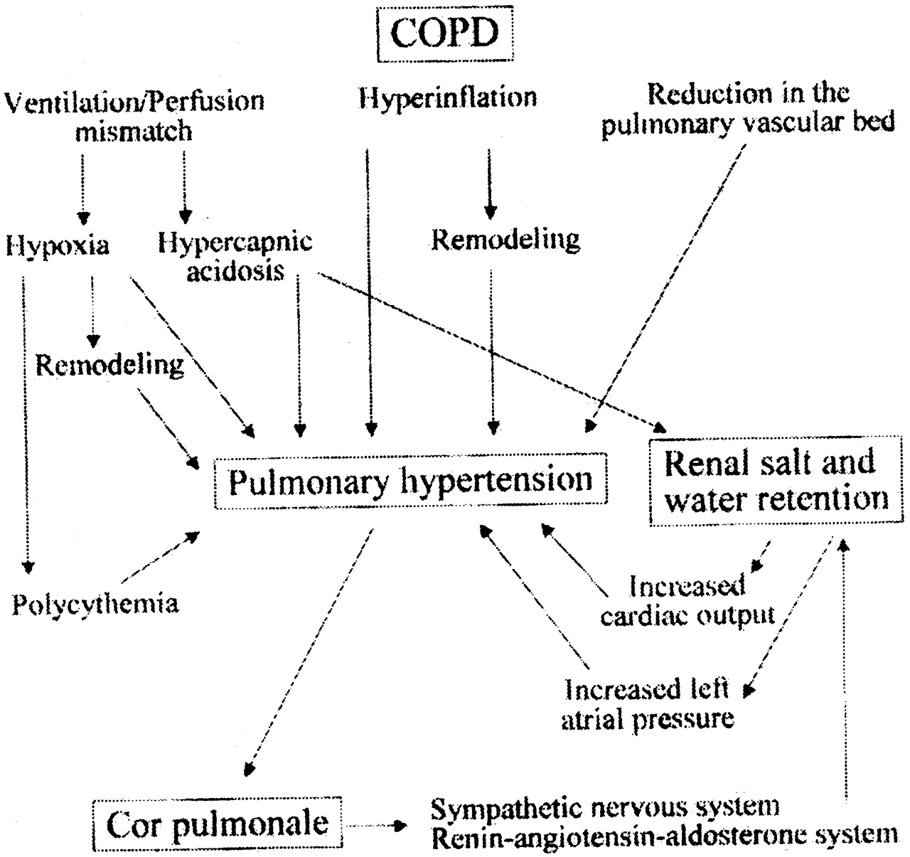
RIGHT VENTRICULAR FAILURE IN COPD


Severe pulmonary hypertension increases right ventricular afterload and eventually leads to the clinical syndrome of right heart failure with systemic congestion and inability to adapt right ventricular output to peripheral demand at exercise. Many patients with advanced COPD present with ankle edema but normal right atrial pressures (at rest). This apparent paradox has stimulated speculation that edema in COPD might be a renal rather than a right ventricular problem .


However, it is now better realized that edema in COPD is likely to be initially caused by repeated stretching of the right atrium from increased right ventricular diastolic pressures at exercise or conceivably with oxygen desaturation during sleep, causing increased sympathetic nervous system tone and activation of the renin-angiotensin-aldosterone system, with resultant renal salt and water retention.
 Renal salt and water retention may be aggravated by hypercapnia, which directly increases proximal tubular reabsorption of sodium, but also activates the sympathetic nervous system and the renin-angiotensin-aldosterone system, which causes additional distal tubular sodium reabsorption through amiloride-sensitive sodium channels.

In summary, and as summarized in , systemic congestion in COPD is caused by right heart failure, involving mechanisms that are very similar to those accounting for systemic and pulmonary congestion in left heart failure, but with an important additional contribution of hypercapnia .

Monday, 22 November 2010

Chest X-rays of the Kyphotic Patient

The radiographer has, by force of habit, angled the X-ray tube caudally for the AP sitting projection. This is a positioning trap in bedside radiography of kyphotic patients. The result is extreme foreshortening of the chest. Even the use of a horizontal ray would have resulted in significant foreshortening of the lung fields (see the lateral image).

This is a lateral chest image taken on a patient with a moderately severe thoracic kyphosis. A triangular positioning sponge has been placed behind the patient. Note that the patient's chest is angled forward considerably and this position is associated with the patient's thoracic kyphosis.

Thursday, 18 November 2010

In lungs: good side down

If a patient has significant right-sided lung disease, his or her preferred recumbent position would be which of the following? Why?
  • a.Right lateral decubitus position
  • b.Left lateral decubitus position
  • c.Prone position
  • d.Supine position
  • e.No preferred position

 The answer is ( b ).
The rule for the comfortable recumbent position in lung disease is “good side down.” This patient with right-sided lung disease will be lying in the left lateral decubitus position to maximize gas exchange in the good lung.
Above is a chest x-ray of a person with Pneumonia. The white arrows on the x-ray are pointing to inflammation in the lung due to germs and mucus.

Wednesday, 10 November 2010

X-ray of a surgically removed lung

Why is this hemithorax opaque?


  • a. There is a large left pleural effusion

  • b. There is a large right pneumothorax

  • c. Atelectasis of the left lung because of a mucus plug

  • d. Pneumonia in the left lung

  • e. The left lung has been surgically removed


Correct Answer: e.The left lung has been surgically removed

Explanation
There is opacification of the left hemithorax. There is a shift of the heart and mediastinal structures (i.e. the trachea) toward the side of opacification. This eliminates pleural effusion, pneumonia and pulmonary edema as possibilities. A tension pneumothorax will displace the heart and mediastinal structures away from the side of the pneumothorax but it will not cause complete opacification of the opposite lung. In addition, there should be no lung markings visible in the hemithorax which contains the pneumothorax. If you look carefully, there is a missing rib on the left side. That indicates a thoracotomy has been performed, at which time the left lung was removed (pneumonectomy) for cancer treatment.


Tuesday, 9 November 2010

Mnemonic for Indications of Bronchoscopy in a child

Indications for Bronchoscopy in Upper Airway Problems in Children use the mnemonic
"SPECS-R"

S - severity of the airway obstruction - Parents subjective impression of the severity of the problem.
P - progression of the obstruction.
E - eating or feeding difficulties.
C - cyanosis.
S - sleep apnea, especially with evidence of cor pulmonale.
R - radiologic abnormalities

See also this video

A PEANUT IN THE RESPIRATORY TRACT CAN CAUSE HUGE PROBLEMS.. WHEN IT IS NEGLECTED FOR WEEKS .... EVEN GREATER PROBLEMS