Thursday, 27 January 2011

ECG changes in acute MI

The ECG changes are often  critical in the diagnosis of acute MI and guiding therapy.
There is a series of ECG changes reflect the evolution of the infarction (The Figure below).
1.The earliest changes are : tall, positive, hyper acute T waves in the ischemic vascular territory.
2.This is followed by elevation of the ST segments (myocardial “injury pattern”).
3.Over hours to days, T-wave inversion frequently develops.
4.And finally, diminished R-wave amplitude or Q waves occur, representing significant myocardial necrosis and replacement by scar tissue, and they are what one seeks to prevent in treating the acute MI .
 Temporal evolution of ECG changes in acute myocardial infarction.
Note the tall hyperacute T waves and loss of R-wave amplitude,followed by ST-segment elevation,T-wave inversion,and development of Q waves.Persistent ST-segment elevation suggests left ventricular aneurysm.

Wednesday, 26 January 2011

Von Hippel-Lindau

Von Hippel-Lindau disease is an autosomal dominant disorder that consists of some combination of retinal angioma, cerebellar or spinal cord hemangioblastoma, renal cell carcinoma, pheochromocytoma, and visceral cysts. About 25% of patients with retinal angiomas will have at least one non-ocular manifestation. About 60% of patients with non-ocular manifestations will have retinal angiomas.

Thus any patient who has Von Hippel-Lindau disease must undergo periodic ophthalmologic examination. Conversely, any patient found to have a retinal angioma should be evaluated for non-ocular manifestations.

The yellow mound fed by a large arteriole and drained by an even larger vein is a capillary hemangioma. It is located in the retinal periphery, beyond the view of the direct ophthalmoscope.

Patients often have many such lesions. If untreated, they will gradually grow, bleed, and leak serous fluid that will finally detach the retina.

Fortunately, laser photocoagulation destroys small lesions. Larger lesions are harder to extinguish; they are treated with a freezing probe placed on the outside of the globe.

Hordeolum Vs Chalazion ...Clinical Summary

A hordeolum is an acute inflammation and/or abscess involving the glandular structures of the eyelid and presents as an acute, tender swelling of the eyelid or eyelid margin. An internal hordeolum involves the meibomian glands, while an external hordeolum (stye) involves the glands of Zeis or Moll at the base of the eye lashes. When bacterial etiology is present, S aureus is the most frequently implicated pathogen.
A chalazion is a firm, nontender lump that arises from obstruction of a meibomian gland. Tenderness, when present, occurs during lid swelling and cyst formation. Both hordeola and chalazia are associated with blepharitis, inflammation of the lid margins associated with plugging of the glandular ducts, and crusting around the lashes. Conditions such as seborrheic dermatitis of the eyelids, rosacea, or rarely eyelash infestation with lice can cause blepharitis and resultant blockage of the meibomian glands.

 Hordeolum. Focal swelling and erythema at the lid margin are seen in this hordeolum.

Chalazion. This chalazion shows nodular focal swelling and erythema from meibomian cyst formation. Pain is present during swelling and cyst formation.

Monday, 24 January 2011

The 5 W's of post-operative fever


Wind---pneumonia, atelectasis at 1st 24- 48 hours
Water---urinary tract infection at Anytime after post op day 3
Wound---wound infections at Anytime after post op day 5
Wonderdrugs---especially anesthesia
Walking---walking can help reduce deep vein thromboses and pulmonary embolus usually occures at Day 7-10

See this video :

Sunday, 23 January 2011

The Supraclavicular Block

About management of cauda equine syndrome

A 45-year-old man complains of decreased sensation in his buttocks and inability to achieve an erection. On examination he has decreased anal sphincter tone and decreased ankle reflexes bilaterally. the best next step in management?
  • A.Bedrest and follow-up in 4 to 6 weeks
  • B.Plain film X-ray of lumbosacral spine
  • C.Sedimentation rate and complete blood count
  • D.Immediate referral for surgical decompression

Answer is D. 

This individual has cauda equine syndrome, and requires immediate surgical decompression to avoid long-term nerve denervation and incontinence/lower extremity weakness. The decreased anal sphincter tone and decreased ankle reflexes indicate a peripheral neuropathy.

Bedrest with follow-up is indicated when no “red flag” symptoms and signs are present.

The plain film X-ray is often normal in patients with cauda equine syndrome.

Koplik’s spots as described

This was a patient who presented with Koplik’s spots on palate due to pre-eruptive measles on day 3 of the illness. Measles is an acute, highly communicable viral disease with fever, conjunctivitis, coryza, cough, and Koplik spots. Koplik spots are ..............

What is Periosteal reaction? it`s types? And how it is helpful in diagnosis.

Periosteal reaction:
A periosteal reaction is a non-specific reaction and will occur whenever the periosteum is irritated by a malignant tumor, benign tumor, infection or trauma.
There are two patterns of periosteal reaction: a benign and an aggressive type.
The benign type is seen in benign lesions such as benign tumors and following trauma.
An aggressive type is seen in malignant tumors, but also in benign lesions with aggressive behavior, such as infections and eosinophilic granuloma.

Benign periosteal reaction
Detecting a benign periosteal reaction may be very helpful, since malignant lesions never cause a benign periosteal reaction.
A benign type of periosteal reaction is a thick, wavy and uniform callus formation resulting from chronic irritation.
In the case of benign, slowly growing lesions, the periosteum has time to lay down thick new bone and remodel it into a more normal-appearing cortex.

Aggressive periosteal reaction
This type of periostitis is multilayered, lamellated or demonstrates bone formation perpendicular to the cortical bone.
It may be spiculated and interrupted - sometimes there is a Codman's triangle.
A Codman's triangle refers to an elevation of the periosteum away from the cortex, forming an angle where the elevated periosteum and bone come together.
In aggressive periostitis the periosteum does not have time to consolidate.


In these X-Rays:

* left:
Osteosarcoma with interrupted periosteal rection and Codman's triangle proximally.
There is periosteal bone formation perpendicular to the cortical bone and extensive bony matrix formation by the tumor itself.
* middle:
Ewing sarcoma with lamellated and focally interrupted periosteal reaction. (blue arrows)
* right:
Infection with a multilayered periosteal reaction.
Notice that the periostitis is aggressive, but not as aggressive as in the other two cases.

Note that: Fibrous dysplasia, Enchondroma, NOF and SBC are common bone lesions.
They will not present with a periosteal reaction unless there is a fracture.
If no fracture is present, these bone tumors can be excluded.
So, Periosteal reaction excludes the diagnosis of Fibrous dysplasia, Enchondroma, NOF and SBC unless there is a fracture

STOP Sepsis Bundle


STOP Sepsis(Strategy to Timely Obviate the Progression of Sepsis) Bundle

CLICK on image for better view

The Physiology of Stress

The Stress Response:
In stress (as in threatening situations that we are unable to cope with) messages are carried along neurones from the cerebral cortex (where the thought processes occur) and the limbic system to the Hypothalamus.

The Anterior Hypothalamus produces sympathetic arousal of the Autonomic Nervous System (ANS). The ANS is an automatic system that controls the heart, lungs, stomach, blood vessels and glands. Due to its action we do not need to make any conscious effort to regulate our breathing or heart beat. The ANS consists of : the sympathetic nervous system and the parasympathetic nervous system. Essentially, the parasympathetic nervous system conserves energy levels. It increases bodily secretions such as tears, gastric acids, mucus and saliva which help to defend the body and help digestion. Chemically, the parasympathetic system sends its messages by a neurotransmitter acetylcholine which is stored at nerve endings.

Unlike the parasympathetic nervous system which aids relaxation, the sympathetic nervous system prepares the body for action. In a stressful situation, it quickly does the following:

* Increases strength of skeletal muscles
* Decreases blood clotting time
* Increases heart rate
* Increases sugar and fat levels
* Reduces intestinal movement
* Inhibits tears, digestive secretions.
* Relaxes the bladder
* Dilates pupils
* Increases perspiration
* Increases mental activity
* Inhibits erection/vaginal lubrication
* Constricts most blood vessels but dilates those in heart/leg/arm muscles

The main sympathetic neurotransmitter is noradrenaline which is released a the nerve endings. The stress response also includes the activity of the adrenal, pituitary and thyroid glands.

The two adrenal glands are located one on top of each kidney. the adrenal medulla is connected to the sympathetic nervous system by nerves. Once the latter system is in action it instructs the adrenal medulla to produce adrenaline and noradrenaline (catecholamines) which are released into the blood supply. The adrenaline prepares the body for flight and the noradrenaline prepares the body for fight. They increase both the heart rate, and the pressure at which the blood leaves the heart; they dilate bronchial passages and dilate coronary arteries; skin blood vessels constrict and there is an increase in metabolic rate. Also gastrointestinal system activity reduces which leads to a sensation of butterflies in the stomach.

Lying close to the hypothalamus in the brain the pituitary gland. In a stressful situation, the anterior hypothalamus activates the pituitary. The pituitary releases adrenocorticotrophic hormone (ACTH) into the blood which then activates the outer part of the adrenal gland, the adrenal cortex. This then synthesises cortisol which increases arterial blood pressure, mobilises fats and glucose from the adipose (fat) tissues, reduces allergic reactions, reduces inflammation and can decrease lymphocytes that are involved in dealing with invading particles or bacteria. Consequently, increased cortisol levels over a prolonged period of time lowers the efficiency of the immune system. The adrenal cortex releases aldosterone which increases blood volume and subsequently blood pressure. Unfortunately, prolonged arousal over a period of time due to stress can lead to essential hypertension.

The pituitary also releases thyroid stimulating hormone which stimulates the thyroid gland, to secrete thyroxin. Thyroxin increases the metabolic rate, raises blood sugar levels, increases respiration/heart rate/blood pressure/and intestinal motility. Increased intestinal motility can lead to diarrhoea. (It is worth noting that an over-active thyroid gland under normal circumstances can be a major contributory factor in anxiety attacks. This would normally require medication.)

The pituitary also releases oxytocin and vasopressin which contract smooth muscles such as the blood vessels. Oxytocin causes contraction of the uterus. Vasopressin increases the permeability of the vessels to water therefore increasing blood pressure. It can lead to contraction of the intestinal musculature.

However, for many people they perceive everyday of their life as stressful. Unfortunately, the prolonged effect of the stress response is that the body's immune system is lowered and blood pressure is raised which may lead to essential hypertension and headaches. The adrenal gland may malfunction which can result in tiredness with the muscles feeling weak; digestive difficulties with a craving for sweet, starchy food; dizziness; and disturbances of sleep.

Thursday, 20 January 2011

Arthritis where the spleen can be FELTY

Felty's syndrome is defined as A combination of rheumatoid arthritis, splenomegaly, leukopenia, pigmented spots on lower extremities, and other evidence of hypersplenism (anemia and thrombocytopenia).
It was first described in 1924
You can remember Felty's syndrome by this :
the arthritis in which the spleen can be felty

It is a potentially serious condition that is associated with seropositive (rheumatoid factor [RF]–positive) rheumatoid arthritis (RA).In which many patients are asymptomatic, some develop serious and life-threatening infections secondary to granulocytopenia.

Tuesday, 18 January 2011

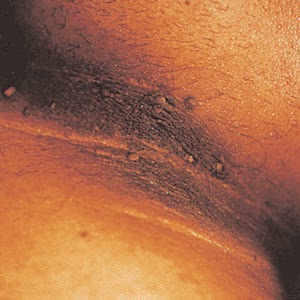
associated diseases with types of Acanthosis nigricans

Which of the following malignancies is associated with the skin condition in the photo?
  • A) Ovarian carcinoma
  • B) Gastric carcinoma
  • C) Malignant melanoma
  • D) Multiple myeloma
  • E) Hodgkin's lymphoma

The answer is B. (Acanthosis nigricans)
Acanthosis nigricans is associated with hyperpigmented areas that typically affect flexural folds (axilla). The two basic types of acanthosis nigricans are benign and malignant.
-The benign form is associated with obesity, diabetes, Stein-Leventhal syndrome, Cushing's disease, Addison's disease, pituitary disorders, and hyperandrogenic syndromes. Drugs, including glucocorticoids, nicotinic acid, diethylstilbestrol, and growth hormone therapy, have also caused acanthosis nigricans. Many cases are idiopathic.
-Malignant acanthosis nigricans is associated with an intestinal cancer such as gastric carcinoma.

Friday, 14 January 2011

Basosquamous Carcinoma in the Temple


This photo shows  large, crusted tumor on the temple that has clinical features of both squamous cell carcinoma (scaly crust), and basal cell carcinoma (translucency in some areas). Histologically, it also has features of both, hence the name basosquamous.

These lesions have a biological aggressiveness intermediate between basal cell and squamous cell carcinomas. They are also called keratotic basal cell carcinoma, squamous cell carcinoma with basaloid differentiation, or "collision tumors."

Tuesday, 11 January 2011

Wolfflin Spots

Wolfflin spots are the whitish clumps of connective fibers found in some irides (mostly blue) near the periphery. These are somewhat distinct from Brushfield spots.
Wolfflin described it as elevated nodules (witish or light yellow ), usually situated in the periphery of the iris, and occuring in about 10% of all normal individual.

Friday, 7 January 2011

How to appreciate Traumatic exophthalmos

Traumatic exophthalmos develops as a retrobulbar hematoma that pushes the eyeball globe outward. Patient presents with periorbital edema, ecchymosis, a marked decrease in visual acuity, and an afferent pupillary defect in the involved eye.
Sometimes the exophthalmos may be obscured by periorbital edema, In this time it can be better appreciated from a superior view. Visual acuity may be affected by the direct trauma to the eye (retinal detachment, hyphema, globe rupture), compression of the retinal artery, or neuropraxia of the optic nerve. The term "Orbital compartment syndrome" occurs when intraorbital pressure exceeds central retinal artery pressure and ocular ischemia ensues. Causes are many with retrobulbar hematoma being the most common.

CT is the best modality to determine both the presence and extent of a retrobulbar hematoma and associated facial or orbital fractures. Consultation of ENT and ophthalmology is indicated on an urgent basis. An emergent lateral canthotomy decompresses the orbit and can be performed in the emergency department and may be sight-saving.
Traumatic Exophthalmos. Blunt trauma resulting in periorbital edema and ecchymosis, which obscures the exophthalmos in this patient. The exophthalmos is not obvious in the AP view and can therefore be initially unappreciated. The Figure below shows the same patient viewed in the coronal plane from over the forehead.
Traumatic Exophthalmos. Superior view, demonstrating the right-sided exophthalmos.

Bone Marrow Aspiration

Thursday, 6 January 2011

How to get a jersey finger !!

Which finger is most likely to be affected with disruption of the flexor digitorum profundus tendon (also known as a jersey finger)?
  • A) Thumb
  • B) Index finger
  • C) Third finger
  • D) Ring finger
  • E) Fifth finger (“pinky”)

Answer and Discussion

The answer is D.

Disruption of the flexor digitorum profundus tendon, also known as jersey finger, commonly occurs when an athlete's finger catches on another player's clothing, usually while playing a tackling sport such as football or rugby.
The injury causes forced extension of the DIP joint during active flexion. The ring finger is the weakest finger and accounts for 75% of jersey finger cases.

Bronchiectasis in High-Resolution CT

Bronchiectasis is defined as localized, irreversible dilatation of part of the bronchial tree. Involved bronchi are dilated, inflamed, and easily collapsible, resulting in airflow obstruction and impaired clearance of secretions. Bronchiectasis is associated with a wide range of disorders, but it usually results from necrotizing bacterial infections, such as infections caused by the Staphylococcus or Klebsiella species or Bordetella pertussis.
Diagnosis of bronchiectasis is based on a clinical history of daily viscid sputum production and characteristic computed tomography (CT) scan findings.

Chest radiography is usually the first imaging examination, but the findings are often nonspecific and the images may appear normal. High-resolution computed tomography (HRCT) scanning has become the imaging modality of choice for demonstrating or ruling out bronchiectasis and its extent.HRCT scanning also helps clinicians to evaluate the status of the surrounding lung tissue and exclude other lesions such as neoplasms.


This is a Transverse high-resolution CT scans obtained in a 40-year-old man with bronchiectasis. (a) Scan shows small-airway disease denoted by centrilobular and tree-in-bud (black arrows) opacities and bronchiolectasis in the left upper lobe. Bronchiectasis in the upper lobe was assigned a grade of 1, with grade 1 (white arrows) bronchial wall thickening. In the apical segments of the lower lobes, grade 2 (arrowheads) bronchial wall thickening also is present. (b) Scan shows a combination of grade 1 (arrows) and 2 (arrowheads) bronchial wall thickening in the basal segments of the lower lobes with an overall bronchial wall thickening score of 1.5. The extent of bronchiectasis was evaluated as grade 3 in the right lower lobe and grade 2 in the left lower lobe. (c) Scan shows mosaic attenuation in both upper lobes. (d) Expiratory scan shows air trapping. The hypoattenuating areas (∗) were confirmed to be caused by air trapping in d.

To know grades of bronchial wall thickening:

Types of Coarctation of the aorta

Infantile type :- aortic stenosis proximal to insertion of ductus arteriosus (preductal).
Adult type :- stenosis is distal to ductus arteriosus (postductal). Associated with notching of the ribs (due to collateral circulation), hypertension in upper extremities, weak pulses in lower extremities,
Associated wilh Turner's syndrome.
Can result in aortic regurgitation.
Check femoral pulses on physical exam.
SO ;
INfantile: IN close to the heart.
ADult: Distal to Ductus. Most commonly associated with bicuspid aortic valve.

Wednesday, 5 January 2011

Craniotomy for resection of 4th Ventricle Tumor

Dr. Pakzaban demonstrates the technique of suboccipital craniotomy for access to the posterior cranial fossa using an Aesculap bone scalpel. This approach is used in many different operations including decompression of Chiari malformation. In this case, it is being used for resection of a 4th ventricle tumor known as subependymoma.

Indications for OPG requests

An OPG or "ORTHOPANTOGRAM", gives a panoramic view of the mandible and teeth.
It is performed using a technique called "tomography". The X-ray tube moves around the head, the x-ray film moves in the opposite direction behind your head. This generates an image slice where the mandible and teeth are in focus, and the other structures are blurred.

Why to get it ?
Dental Disease
* Caries - appear as different shaped areas of radiolucency in the crowns or necks of teeth.
* Peridontioiditis - when inflammation extends into the underlying alveolar bone and there is a loss of attachment.
* Peridontal Abscess - Radiolucent area surrounding the roots of the teeth.


Extraction of teeth (eg. wisdom teeth)
* OPG shows angulation, shape of roots, size and shape of crown, effect on other teeth.


Teeth Abnormalities
* Eg. Developmental, to show size, number, shape and position.


Trauma to teeth and facial skeleton
* Mandible fractures are often bilateral.
* Panoramic view of mandible to view the fracture.
* Determine site and direction of fracture lines.
* Relationship of teeth to fracture lines.
* Alignment of bone fragments after healing.
* Evidence of infection or other complications post intervention.
* Follow up to assess healing.


Transplant workup
* To look for evidence of any underlying dental disease (eg. abscess)
* Patients on steriods after a transplant are immunosuppressed and the mouth is a common site of infection.

Different arterial pulse waveforms and example causes

This is best assessed at the carotid artery. You are feeling for the speed at which the artery expands and collapses and force with which it does so. It takes some practise to master and it may be useful to imagine a graph such as those shown in the figure below,also Some examples are present:

Aortic stenosis: a slow rising pulse, maybe with a palpable shudder. Sometimes called anacrotic or a plateau phase.
Aortic regurgitation: a collapsing pulse which feels as though it suddenly hits your fingers and falls away just as quickly. You could try feeling at the brachial artery and raising the arm above the patient's heart. Sometimes referred to as a waterhammer pulse.
Pulsus bisferiens: a waveform with 2 peaks, found where aortic stenosis and regurgitation co-exist.
Hypertrophic cardiomyopathy: this pulse may feel normal at first but peter out quickly. Often described as jerky.
Pulsus alternans: an alternating strong and weak pulsation, synonymous with a severely impaired left ventricle in a failing heart.
Pulsus paradoxus: pulse is weaker during inspiration (causes include cardiac tamponade, status asthmaticus, and constrictive pericarditis).


Graphical representation of different arterial pulse waveforms and their causes

Tuesday, 4 January 2011

Le Fort Classification of facial fractures

In 1901 ;René Le Fort (1869-1951),French surgeon, reported his work on cadaver skulls that were subjected to blunt forces of various magnitudes and directions. He concluded that predictable patterns of fractures follow certain types of injuries. Three predominant types were described.


Le Fort I fractures (horizontal) may result from a force of injury directed low on the maxillary alveolar rim in a downward direction. The fracture extends from the nasal septum to the lateral pyriform rims, travels horizontally above the teeth apices, crosses below the zygomaticomaxillary junction, and traverses the pterygomaxillary junction to interrupt the pterygoid plates.

Le Fort II fractures (pyramidal) may result from a blow to the lower or mid maxilla. Such a fracture has a pyramidal shape and extends from the nasal bridge at or below the nasofrontal suture through the frontal processes of the maxilla, inferolaterally through the lacrimal bones and inferior orbital floor and rim through or near the inferior orbital foramen, and inferiorly through the anterior wall of the maxillary sinus; it then travels under the zygoma, across the pterygomaxillary fissure, and through the pterygoid plates.

Le Fort III fractures (transverse), also termed craniofacial dysjunctions, may follow impact to the nasal bridge or upper maxilla. These fractures start at the nasofrontal and frontomaxillary sutures and extend posteriorly along the medial wall of the orbit through the nasolacrimal groove and ethmoid bones. The thicker sphenoid bone posteriorly usually prevents continuation of the fracture into the optic canal. Instead, the fracture continues along the floor of the orbit along the inferior orbital fissure and continues superolaterally through the lateral orbital wall, through the zygomaticofrontal junction and the zygomatic arch. Intranasally, a branch of the fracture extends through the base of the perpendicular plate of the ethmoid, through the vomer, and through the interface of the pterygoid plates to the base of the sphenoid.

USMLE Pass Program Video Lectures (2010) - Pediatrics

About the PASS Program:

The PASS Program was created in 1988 and officially opened for business in the year 2000. This program is different from any other program in this country. It teaches integrated thinking so that a medical student can answer any question asked at any level. It does not, and should not, matter whether a student is taking the USMLE 1, USMLE 2, or USMLE3. The information that is covered in this program is so high yield and so highly integrated that it is relevant on all exams, even board certification exams. Because of this unique format, we have been able to help students achieve the highest pass rates, as well as the highest passing scores, on all of the USMLE exams.

The mission of the PASS PROGRAM:

The mission of the PASS PROGRAM is to inspire, encourage and empower medical students and physicians who enter our doors. We will not only teach them the basic medical sciences, but we will teach them how to clinically integrate their knowledge. In other words, we will encourage them not to merely memorizemedical information , but empower them to clearly and clinically master that knowledge. We will maintain a positive learning environment at all times so that no student will feel ashamed or be embarrassed of his or her academic situation. For, in the end, our true mission is to show other medical institutions that even when a student may not look so promising with a poor academic history, with the individualized teaching sessions we have with all of our students, most of them can be helped to develop into academically strong, spiritually empowered, and outstandingly confident clinical physicians. With God as our inspiration, this will always be our mission.

The clarity and simplication of the explanations of certain key concepts are second to none. You will understand and retain the specificities of the pathophysiology of the most high yield disease processes that you will be tested on. You will also be provided with test taking skills that will enable you to quickly recognize distractors and clues. Over 71 hours of Live lecture by Dr. Francis.

CLASS ONE - CLASS TWO - CLASS THREE

Synthesis & Transport of Thyroid Hormones

The thyroid secretes 2 iodine-containing hormones: thyroxine (T4 ) and triiodothyronine (T3). The iodine necessary for the synthesis of these molecules comes from food or iodide supplements. Iodide ion is actively taken up by and highly concentrated in the thyroid gland, where it is converted to elemental iodine by thyroidal peroxidase ( See the Figure ).
The protein thyroglobulin serves as a scaffold for thyroid hormone synthesis. Tyrosine residues in thyroglobulin are iodinated to form monoiodotyrosine (MIT) or diiodotyrosine (DIT) in a process known as iodineorganification.
Within thyroglobulin, 2 molecules of DIT combine to form T4, while 1 molecule each of MIT and DIT combine to form T3. Proteolysis of thyroglobulin liberates the T4 and T3, which are then released from the thyroid. After release from the gland, T4 and T3 are transported in the blood by thyroxine-binding globulin, a protein synthesized in the liver.

This figure also show Sites of action of some antithyroid drugs. I–, iodide ion; I°, elemental iodine. Not shown: radioactive iodine (131I), which destroys the gland through radiation.

Monday, 3 January 2011

Inspecting a skin lesion

 # Inspect each lesion carefully and note¦
-Grouped or solitary? Pattern if grouped .
-Distribution/location:
  • Symmetrical/asymmetrical?
  • Peripheral?
  • In only light exposed areas?
  • Dermatomal?
-Colour.
-Shape.
-Size.
-Surface.
-Edge.
-Nature of the surrounding skin.

Histopathology of Osteosarcoma

The staging evaluation of Esophageal Cancer

After establishing a diagnosis of esophageal cancer, adequate staging is required, because staging is the most important step in choosing appropriate therapy. More than 50% of patients have unresectable or metastatic disease at the time of presentation. For the others, survival is closely related to the stage of the disease.
The staging evaluation allows patients to be assigned a clinical stage according to the American Joint Committee on Cancer tumor-node-metastasis (TNM) classification. Informed recommendations about therapy and appropriate information regarding prognosis depends on this clinical staging, an assessment that can, however, only approximate the true disease stage.
 Primary Tumor (T)

* TX: Primary tumor cannot be assessed
* T0: No evidence of primary tumor
* Tis: Carcinoma in situ
* T1: Tumor invades lamina propria (T1a) or submucosa (T1b)
* T2: Tumor invades muscularis propria
* T3: Tumor invades adventitia
* T4: Tumor invades adjacent structures

Regional Lymph Nodes (N)

* NX: Regional lymph nodes cannot be assessed
* N0: No regional lymph node metastasis
* N1: Regional lymph node metastasis
* N1a: One to three nodes involved
* N1b: Four to seven nodes involved
* N1c: More than seven nodes involved

Distant Metastasis (M)

* MX: Distant metastasis cannot be assessed
* M0: No distant metastasis
* M1: Distant metastasis
1. Tumors of the lower thoracic esophagus:
- M1a: Metastases in celiac lymph nodes
- M1b: Other distant metastases
2. Tumors of the midthoracic esophagus:
- M1a: Not applicable
- M1b: Nonregional lymph nodes and/or other distant metastases
3. Tumors of the upper thoracic esophagus:
- M1a: Metastases in cervical nodes
- M1b: Other distant metastases

Port wine stains and Associations

Port wine stains (PWS) are almost always evident at birth and are a capillary malformation. They are usually sporadic, although some occur in families in an apparently autosomal dominant inheritance. They are initially pink or red, and grow in proportion to infant growth. Although they may appear to lighten in the first few months, they generally darken after this.

They can be associated with other skin anomalies such as extensive mongolian spots. An important association is Sturge-Weber syndrome ( PWS+mental retardation ): PWS involving the forehead (V1 area of the trigeminal nerve), eye abnormalities (choroidal vascular abnormalities, glaucoma), and leptomeningeal and brain abnormalities (vascular malformations, calcification, or cerebral atrophy). Another association is Klippel-Trenaunay syndrome, with a capillary malformation and overgrowth of the soft tissue and bone of the affected limb.

Treatment of PWS is possible using a laser, although improvement rather than complete resolution is most common.

Sunday, 2 January 2011

Chronic Gouty Tophi

After years of gouty attacks, patients will develop a chronic arthritis resulting in bone and cartilage destruction and deformity. Uric acid crystals deposit within and surrounding the joint causing a chronic destructive inflammatory process. X-rays characteristically show well demarcated "punched out" erosions.
Tophi at helix of ear.
 Tophi or deposits of uric acid crystals are often found around joints, in the olecranon bursa or at the pinna of the ear.


X-ray showing soft tissue swelling and erosion of 2nd PIP.

Pneumocephalus

X-ray showing nasal and orbital fractures and pneumocephalus
Pneumocephalus is the presence of air in the cranial vault. It is usually associated with neurosurgery, barotrauma, basilar skull fractures, sinus fractures, nasopharyngeal tumor invasion and meningitis.Headache and altered consciousness are the most common symptoms.
Tension pneumocephalus can occur and is a neurosurgical emergency.Plain X-rays can diagnose pneumocephalus, but CT scan is the diagnostic modality of choice.A classical CT sign of tension pneumocephalus is the “Mount Fuji sign”: the massive accumulation of air that separates and compresses both frontal lobes and mimics the profile of the large volcano in Japan.
Large right pneumocephalus compressing right frontal lobe and widening interhemispheric space. There are also air bubbles in basal cisterns and cerebellar fissures bilaterally

Most cases of pneumocephalus resolve spontaneously, and conservative management should be provided. Nonoperative management involves oxygen therapy, keeping the head of the bed elevated, prophylactic antimicrobial therapy (especially in post-traumatic cases), analgesia, frequent neurologic checks and repeated CT scans.

Supraspinatus Pathology by Ultrasound

This video details the complex structures of the human shoulder and how proper and careful ultrasound scanning techniques can identify tendon and rotator cuff tears, as well as avoid false positive and false negative tear diagnoses.

Leishmania donovani Amastigotes

Leishmania donovani amastigotes in a spleen smear, spleen section and liver section.

Diagram for Effects of sickle cell anemia

Minimally Invasive Robotic Surgery for Hysterectomy

Robotic surgery enables physicians to perform minimally invasive surgery that was not possible with simple laparoscopic instruments. Its one of the greatest advancements in surgery during our lifetime. With the robots high definition camera, 10 times magnification and 540 degrees of movement we are able to perform surgery that could not be accomplished with traditional methods.

Carolina Robotic center is a division of WHA Midcarolina obstetrics and Gynecology P.A.

Saturday, 1 January 2011

Anemias and it`s corresponding RBC`s shape

Erythrocytes may have abnormal shapes and sizes in certain diseases.In iron deficiency you would expect to see ?!
  • a.Microcytic, hypochromatic anemia with smaller mature erythrocytes
  • b.Macrocytic, hyperchromatic anemia with fewer, larger mature erythrocytes
  • c.Poikilocytosis and more fragile erythrocytes
  • d.Spherocytosis
  • e.No change in erythrocyte size or shape, but a substantial drop in the hematocrit

The answer is a.
Iron deficiency leads to anemia with the presence of smaller, pale-staining erythrocytes (microcytic, hypochromatic). Hyperchromatic, macrocytic anemia results from vitamin B 12 deficiency (answers b and e).

The presence of spherical rather than biconcave erythrocytes is known as spherocytosis (answer d). The RBC membrane undergoes deformation due to the inability of ankyrin to bind spectrin. The shape change results in trapping in the splenic sinusoids and excessive destruction of red blood cells in that organ. Poikilocytosis is the generic term for abnormally shaped erythrocytes (answer c).
Hereditary elliptocytosis and hereditary poikilo-cytosis are inherited diseases in which there is RBC membrane fragility and abnormal shape due to spectrin mutations. Mutations in Band 3 (anion exchanger 1) result in RBCs that are hyperchromatic with poikilocytosis.

Contraindications to MRI procedure

The principal contraindications of the MRI procedure are mostly related to the presence of metallic implants in a patient. The risks of MRI scans increase with the used field strength. In general, implants are becoming increasingly MR safe and an individual evaluation is carried out for each case.

Absolute Contraindications for the MRI scan:
# Intracranial aneurysm clips (Unless the referring physician is certain that it is made of nonferromagnetic material such as titanium).

# Intra-orbital metal fragments

# Any electrically, magnetically or mechanically activated implants (including cardiac pacemakers, biostimulators, neurostimulators, cochlear implants, and hearing aids).


Relative Contraindications for the MRI scan:
-cochlear implants
-other pacemakers, e.g. for the carotid sinus
-insulin pumps and nerve stimulators
-lead wires or similar wires (MRI Safety risk)
-prosthetic heart valves (in high fields, if dehiscence is suspected)
-haemostatic clips (body)
-non-ferromagnetic stapedial implants