Recession, Limited Gingival Band, Esthetics and Stability are addressed with this Periodontal Microsurgical procedure using a purified collagen implant: AlloDerm

health, acne, acne vulgaris, cancer,
Showing posts with label DENTISTRY. Show all posts
Showing posts with label DENTISTRY. Show all posts
Sunday, 27 February 2011
Wednesday, 5 January 2011
Indications for OPG requests

It is performed using a technique called "tomography". The X-ray tube moves around the head, the x-ray film moves in the opposite direction behind your head. This generates an image slice where the mandible and teeth are in focus, and the other structures are blurred.
Why to get it ?
Dental Disease
* Caries - appear as different shaped areas of radiolucency in the crowns or necks of teeth.* Peridontioiditis - when inflammation extends into the underlying alveolar bone and there is a loss of attachment.
* Peridontal Abscess - Radiolucent area surrounding the roots of the teeth.
Extraction of teeth (eg. wisdom teeth)
* OPG shows angulation, shape of roots, size and shape of crown, effect on other teeth.Teeth Abnormalities
* Eg. Developmental, to show size, number, shape and position.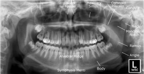
Trauma to teeth and facial skeleton
* Mandible fractures are often bilateral.* Panoramic view of mandible to view the fracture.
* Determine site and direction of fracture lines.
* Relationship of teeth to fracture lines.
* Alignment of bone fragments after healing.
* Evidence of infection or other complications post intervention.
* Follow up to assess healing.
Transplant workup
* To look for evidence of any underlying dental disease (eg. abscess)* Patients on steriods after a transplant are immunosuppressed and the mouth is a common site of infection.
Friday, 31 December 2010
Indirect IA (Inferior Alveolar) nerve block on a model
University of The Pacific Arthur A. Dugoni School of Dentistry present a video illustrating the technique of Inferior Alveolar Nerve Block on a model by Dr. Anders Nattestad, Professor and Director, Department of Oral and Maxillofacial Surgery.
Monday, 20 December 2010
Dental Implant Surgery (lateral incisor)
Full surgery of a zirconia dental implant. Implant type 12. Upper Left Lateral Incisor. 8 min. Very esthetic. 100% biocompatible. Metal Free. High percentage of osseointegration.
Thursday, 16 December 2010
Anatomical parts of Gingiva

The junction between them is called the mucogingival junction. The small margin of tissue outlined in yellow on the lower diagram is called the free or marginal gingiva (sometimes called the free gingival margin), and it is the unattached, sleevelike portion of the gingiva that encircles the tooth to form the gingival sulcus.
The Vermillion border is the junction of the dry, pink part of the lip with the skin of the face. The labial (lip) vestibule is marked on the diagram. The Upper Labial Frenum is also visible.
Thursday, 4 November 2010
Palato-Pharyngeal Function
Surgical patients with parts of their faces surgically removed show palato-pharyngeal functions.
Subscribe to:
Posts (Atom)